Elevate Your Research Efficiency with MoticFlexScan 60
Automate high-volume low-plex fluorescence whole slide imaging in an open-source tool tailored for any lab.
- Economical high-throughput fluorescence scanning
- Flexibility in transitioning between brightfield and fluorescence imaging optimize research efficiency
- Ideal for high-volume kidney applications
Access slides scanned directly from the MoticFlexScan fluorescence and discover exceptional high-resolution image quality right from your desktop.
We have millions of cases from 70+ years
Use Cases
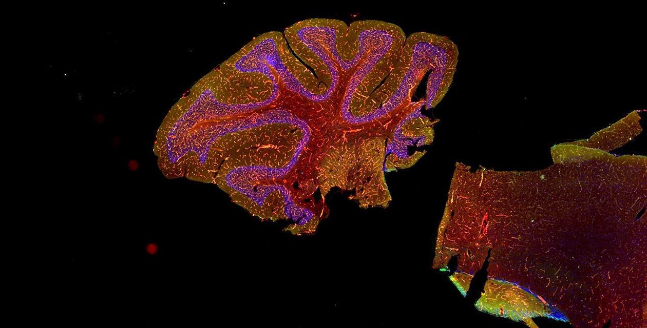
For Kidney
The MoticFlexScan 60 is a leading slide scanner for high-volume kidney tissue samples. It effortlessly handles kidney biopsy specimens stained with up to 10 different markers per case, efficiently scanning serial sections to enable thorough analysis of each digital slide. With support for 3-color, 2-color or single-plex FITC staining, it offers unparalleled flexibility. Its advanced imaging capabilities guarantee outstanding image quality, allowing for meticulous examination of crucial histological features. Furthermore, seamless integration with existing laboratory information systems and third-party image analysis software ensures smooth workflow, efficient data management, collaboration, and reporting.
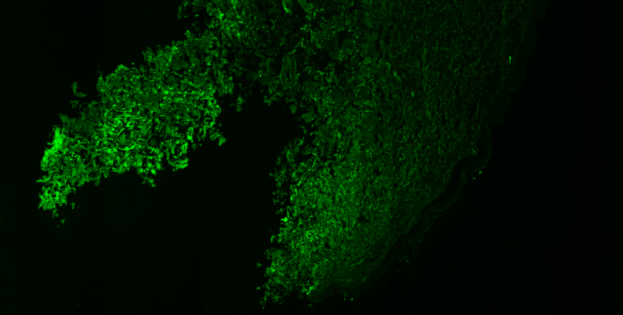
For Skin
The MoticFlexScan 60 is the perfect digital pathology solution for dermatopathology labs. It handles diverse skin tissue samples, even challenging ones. Dermatologists can use two-plex staining protocols with DAPI as a counterstain for comprehensive visualization of epidermal layers, dermal features, and key histological elements. Its fluorescence overview Preview Mode feature optimizes scanning by helping users find fluorescently stained tissue or add focus points as needed. Additionally, it enables switching between brightfield and fluorescence imaging on any slides. This versatility allows dermatopathologists to leverage the most appropriate imaging modality for their specific diagnostic needs, whether it's comprehensive visualization of histological structures or targeted analysis of fluorescent markers.
MoticFlexScan 60
Automate high-volume low-plex fluorescence whole slide imaging in an open-source tool tailored for any lab.
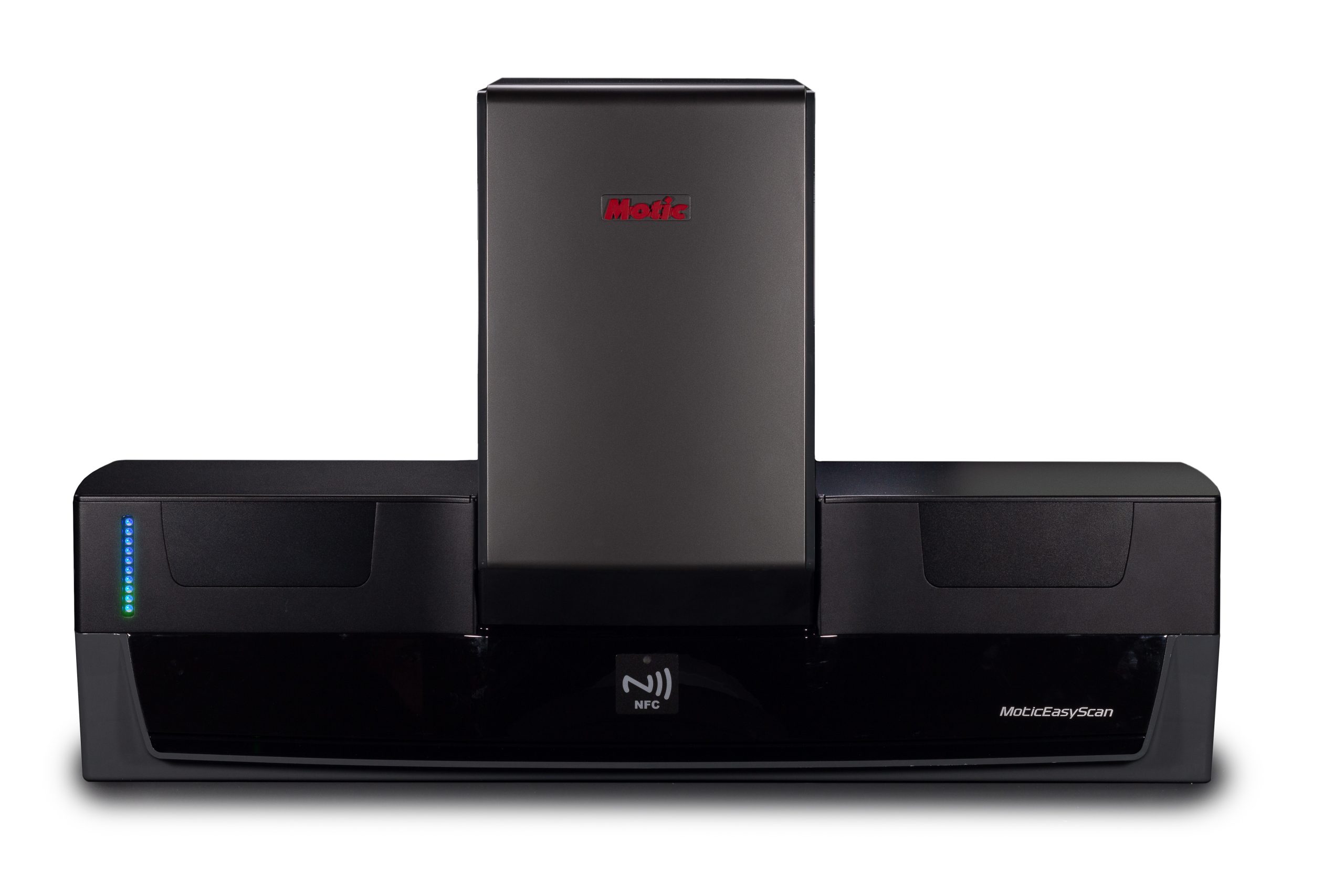
Navigate through tricky samples to automatically detect where on your slide to scan. It also features a Preview Mode designed to find faintly stained tissue and allow the use to fixup or add focus points if needed
Tailored for low-plex, high-volume kidney studies, providing a reliable tool for analyzing numerous whole slide samples.
Streamline your workflow with our convenient perk
Workflow enhancement with
MoticFlexScan 60
The Challenge: Disruptive Workflow for Brightfield and Fluorescence Imaging
Users that use brightfield and fluorescence imaging commonly need to switch between different equipment, causing disruptions in workflow and the inability to do side-by-side comparisons
The Motic Solution: All-in-One Solution for Brightfield and Fluorescence Imaging
The MoticFlexScan 60 holds multiple light sources and can conduct both BF and FL imaging, allowing for easier switching and localized comparisons with a click of a button.
The Challenge: Hindered ROI Identification
Users may spend additional time adjusting settings to locate their ROI, thereby reducing productivity and efficiency.
The Motic Solution: Enhance ROI detection
The MoticFlexScan 60 Preview Mode is designed to detect faintly stained tissue and enable users to adjust or add focus points effortlessly.
The Challenge: Managing Non-Standard File Formats in Digital Pathology Data
Users struggle when they have to use non-standard files for handling digital pathology data. This limitation makes it difficult to share, exchange data, and access expert opinions
The Motic Solution: Flexible file format meeting DICOM standard
MoticFlexScan 60 uses common formats (.tiff, .svs and .png), complies with the DICOM standard and offers open-source formats, allowing for easy integration with various organizational systems and workflows.
See what you get
MoticFlexScan 60 key features benefits
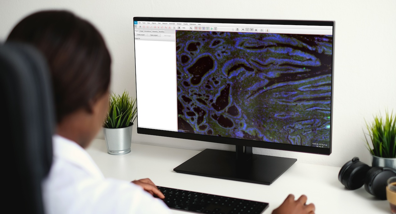
Integration and Interoperability with 3rd party software image analysis tools
MoticFlexScan seamlessly integrates with third-party software image analysis tool like QuPath through open file formats, ensuring a streamlined and efficient analysis process. By embracing interoperability, users can leverage QuPath's advanced image analysis tools alongside MoticFlexScan's high-resolution imaging capabilities, enhancing pathology research and diagnosis. With support for widely-used file formats such as pyramid .TIFF and .QPTIFF, it allows for easy exchange of high-quality whole-slide images between the scanner and third-party analysis tools. This interoperability not only streamlines workflows but also future-proofs digital pathology investments, enabling efficient data management, collaboration, and reporting across the digital pathology ecosystem.
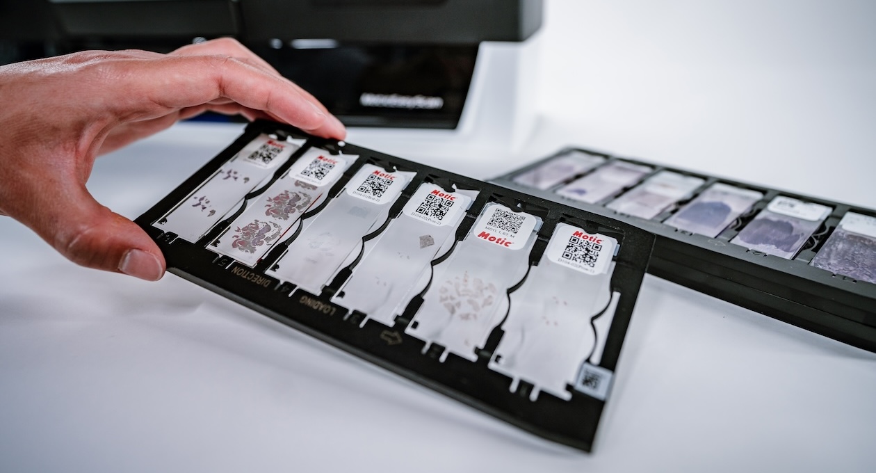
Tray-Centric Design Ensuring Fluorescence Slide Security
MoticFlexScan 60 is engineered with a tray-centric design to safeguard your precious fluorescently stained samples, eliminating the risk of breakage and ensuring project completion. Unlike traditional slide loaders that rely on complex robotic arms to transport the samples, MoticFlexScan 60 keeps your slides stationary within the tray. Designed with fewer moving parts (and no robotic arms) this instrument is more reliable than traditional scanners because the likelihood of mechanical malfunctions that could jeopardize the integrity of your fluorescence-stained samples has been greatly reduced. With MoticFlexScan 60's tray-centric design and limited moving parts, you can trust that your fluorescence slides will be handled with the utmost care, allowing you to focus on the critical task of analyzing your tissue samples.
We offer fully integrated software platforms to continue your digital workflow
Software platform
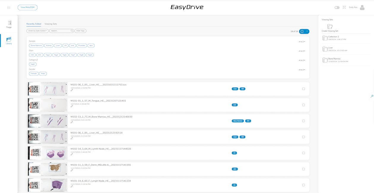
EasyDrive: Organizational Hub for Centralized Data Storage
EasyDrive seamlessly integrates with MoticEasyScan to enhance digital pathology workflows, offering a platform for sharing and managing data. It serves as an organized encyclopedia of digital pathology knowledge, enabling users to curate scanning runs systematically for easy access and sharing. With features like triage, users can effortlessly sort finished projects and isolate unneeded slides, while the library functionality allows for simple organization, search, and retrieval of slides. User can share slides via tiny URL-style link and archive unique samples for future study, ensuring that valuable data is preserved and accessible whenever needed. With MoticEasyScan and EasyDrive integration, laboratories can streamline their workflow, improve organization, and enhance collaboration in digital pathology.

LIS Connect: Customized Integration for Seamless Pathology Connectivity
LIS Connect provides Application Programming Interface (API) and remote viewing tools equipped with barcode parsing capabilities. It is tailored to provide efficient project planning and execution tools, along with scalability to meet requirements from entry-level to enterprise-level needs. With its intuitive user-friendly viewer integration and modern web viewer for pathology, LIS Connect seamlessly enhances your existing Lab Information System (LIS) with digital pathology functionality. By integrating the viewer with the LIS, it enables a smooth connection between electronic patient record-keeping systems and digital pathology software solutions. Additionally, LIS Connect can be optimized with barcode tracking to automate LIS data field population, thereby reducing manual entry errors and enhancing data accessibility and workflow efficiency.
FAQs
It is highly dependent on the fluorescence level of the dyes and how high the exposure level needs to be and the tissue sample.
The below wavelengths are currently in a multi-band filter cube included in the unit
- Blue wavelengths excited between 385-405nm (emission between 425-455)
- Green wavelengths excited between 410-495nm (emission between 505-540)
- Orange wavelengths excited between 550-575nm (emission between 585-625)
The MoticFlexScan can provide images in .qpitff and a variety of different (.svs, .tiff, .png). Qptiff is commonly used as an opensource format and allows for use with analysis software, like QuPath.
Struggling with Your Workflow? Let’s Find a Solution.
Get a Free Consultation—Tailored to Your Needs.

Michael Kasten,
Motic Digital Pathology Expert
I am ready to provide my best support. Ask your question via the contact form or get in touch with me directly.
If you need assistance with your device, please contact Motic's
customer support
Is inefficiency slowing down your lab? Need a better way to manage your slides?
Connect with our experts for a personalized consultation and discover how MoticEasyScan Two can streamline your workflow, improve collaboration, and enhance efficiency.
Get answers within 24 hours and take the first step toward a smarter solution.

