Ready for a scanner that can grow with you?
Discover future-ready small-footprint scanner upgradability with the MoticEasyScan Pro 24
- Industry-first upgradable path to high-volume MoticEasyScan Infinity scanner to keep up with the demand of the future
- Excellent image quality up to 80x (0.13 μm/px)
- Automation meets Frozen-Section Live
View slides scanned directly from the MoticEasyScan Pro 24 and explore high resolution
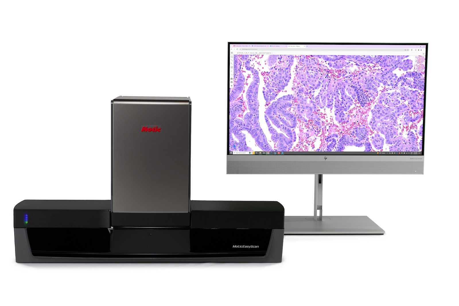
We have millions of cases from 70+ countries
Use Cases
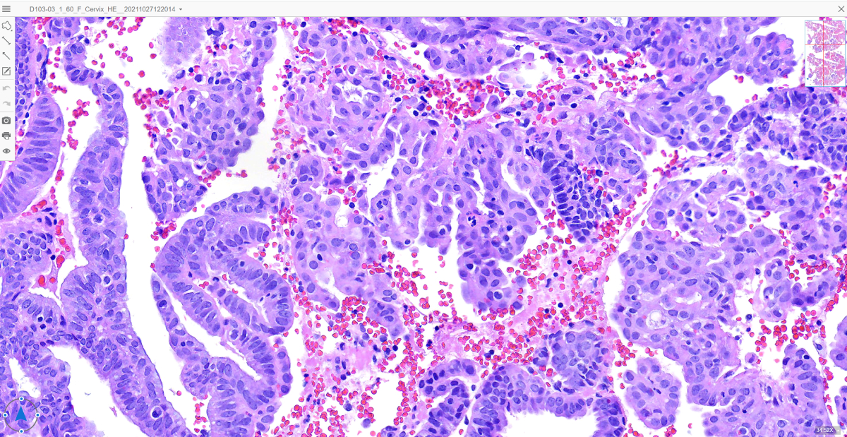
Ideal for H/E sample with IHC serial sections
Specifically designed to enable IHC quantification, designed with Motic optics to aid in accurate interpretation of staining patterns and cellular morphology, making quantifying IHC staining precise and more efficient.
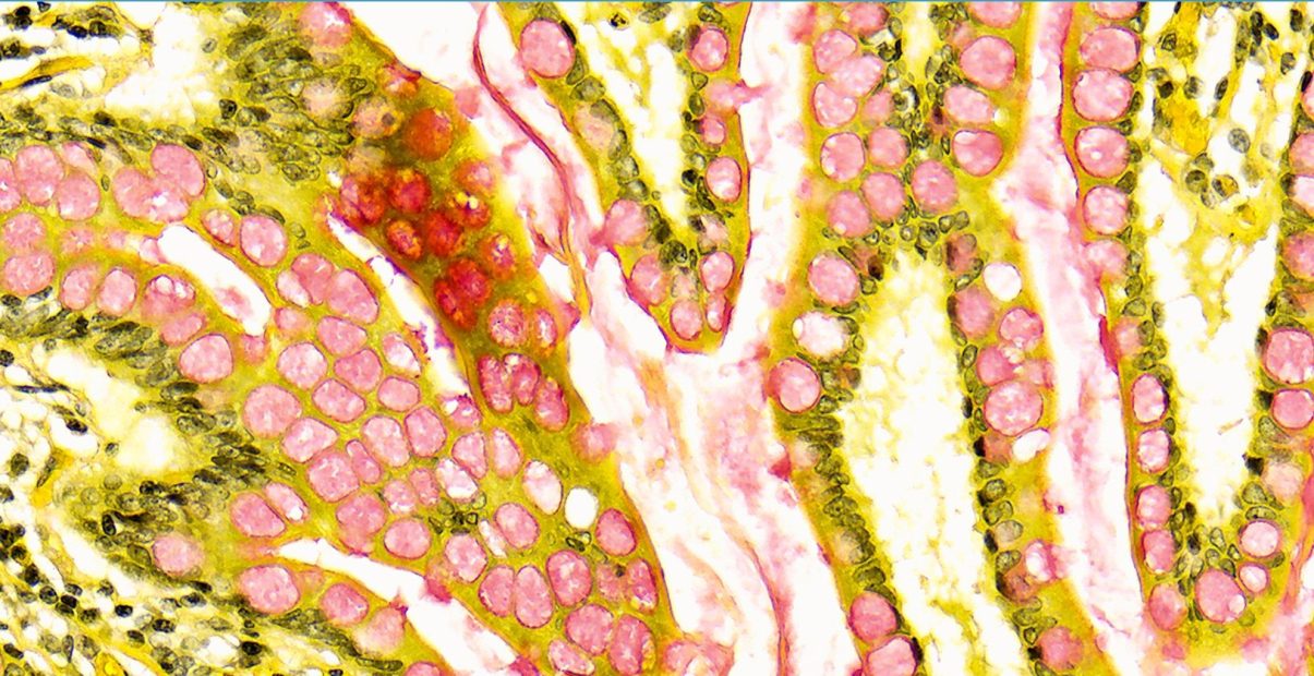
Streamlined Remote Frozen Section Scanning
Optimize workflow with efficient high-volume remote frozen section scanning for timely assessments. Whether connecting multiple labs and sites or enabling rapid collaboration, Frozen Section Live eliminates the need for travel to remote locations and the time-consuming scanning of whole slide images. View and operate slides live with our industry leading Autofocus technology
MoticEasyScan Pro 24
Transform your pathology lab with the versatile MoticEasyScan Pro 24 digital scanner, revolutionizing digital pathology.
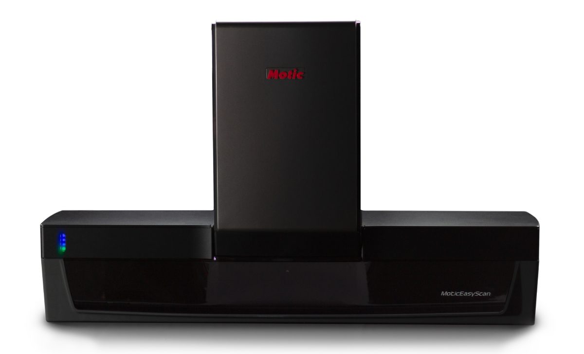
When the time comes, upgrade from 24-slide capacity to 100-slide capacity for an extra productivity lift, ensuring your lab keeps up with the demands of the future.
MoticEasyScan empowers you to navigate through tricky samples with tissue detection algorithms for faintly stained tissue, as well as Preview Mode to enhance your workflow productivity.
A powerhouse for high-volume FS-Live operations, ensuring seamless and efficient scanning for various applications.
Explore Z-Stack and EDF capabilities for flexible whole-slide scanning, accommodating even the thickest samples with ease.
Featuring 12-Megapixel camera for broader view of morphology, capturing intricate details effortlessly.
Utilize a separate focusing camera for quick tissue detection, bypassing lengthy image pre-mapping process for efficient scanning.
Need More Details
MoticEasyScan Pro 24 Specification sheet
Stremline your workflow with our convenient perk
How MoticEasyScan Pro 24 make life easier
Get more projects done
- The 12 megapixel camera provides a broader view of morphology, enabling quicker live-mode reviews
- Ability to preview all samples before scanning
Lightning Fast Frozen Section Live
- View slides continuously in live mode
- Handle larger-scale frozen sections workloads without manual intervention
Scalability for business growth
- Cost-effective solution for handing increased workloads, preventing workflow bottlenecks, and supporting successful business expansion
- Flexibility to meet high demand without the need for investing in a new digital scanner
Improve workflow with minimal set up time
- Continuous loading
- Maximizing throughput with just a fraction of your bench space
Wide-ranging applications with ease
Applications
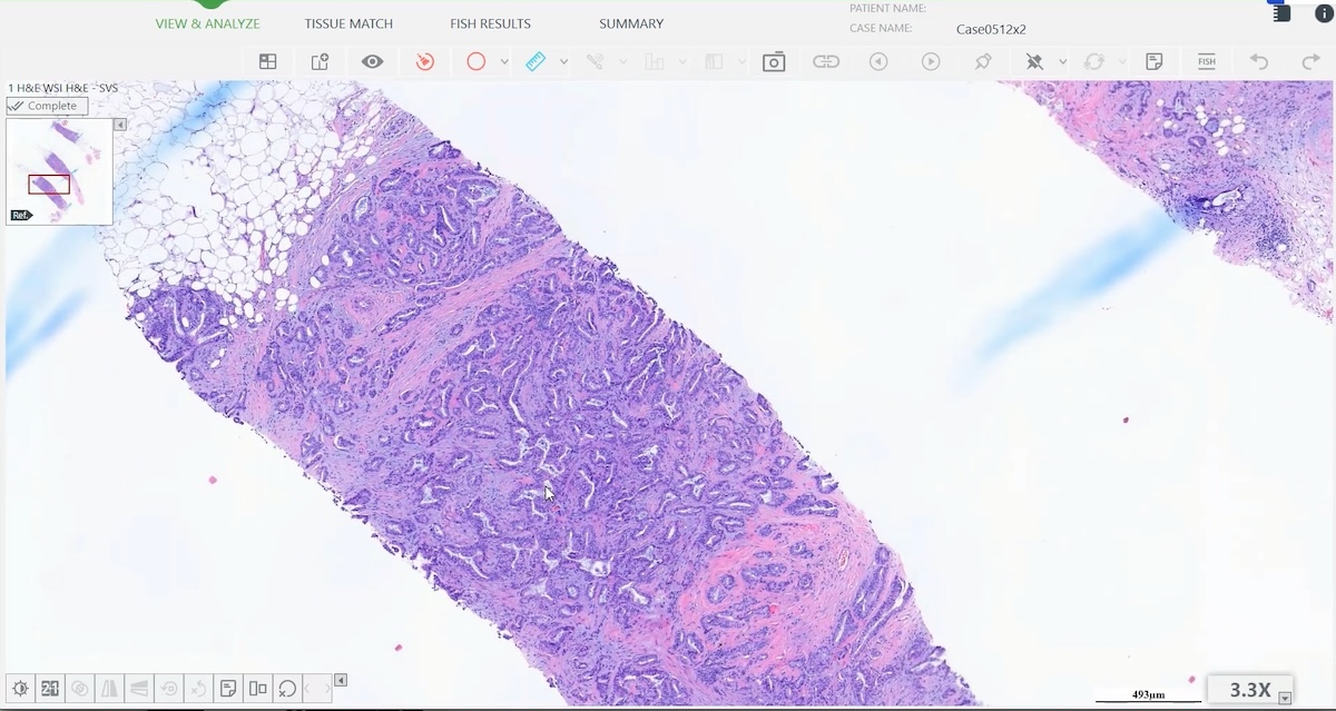
Integration with Image Analysis Software Platform
MoticEasyScan seamlessly integrates with various image analysis software platforms to enhance histopathology practices. The imaging and analysis systems, when paired with MoticEasyScan , cater to a wide range of histopathology needs, including Quantitative IHC Scoring and Whole Slide Imaging of H&E/IHC tools from image analysis leaders, including Applied Spectral Imaging, based on the Motic open-source platform. By leveraging interoperability with most computer-assisted analysis pipelines and staying laser focused on providing exceptional image quality of MoticEasyScan, pathologists can achieve uncompromising standardization in results interpretation, enabling streamlined workflows and improved efficiency in evaluating patient cases.
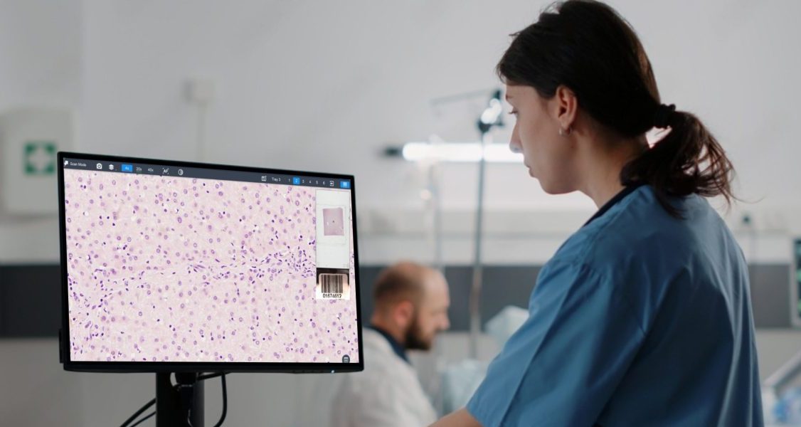
Integration and Interoperability with Frozen Section Live
MoticEasyScan, featuring Frozen Section Live, presents versatile applications across laboratory use, research, and education. In laboratory settings, it streamlines scanning processes for frozen sections, offering real-time visualization that enables swift diagnostic assessments for timely patient care. For researchers, the system's high-quality imaging capabilities support detailed tissue sample analysis, complemented by its compatibility with third party Quantitative IHC Scoring for research investigations. In educational environments, MoticEasyScan fosters interactive learning through real-time slide viewing and remote access, empowering students with hands-on experience and providing educators with tools to enhance collaborative learning opportunities.
We offer fully integrated software platforms to continue your digital workflow
Software platform
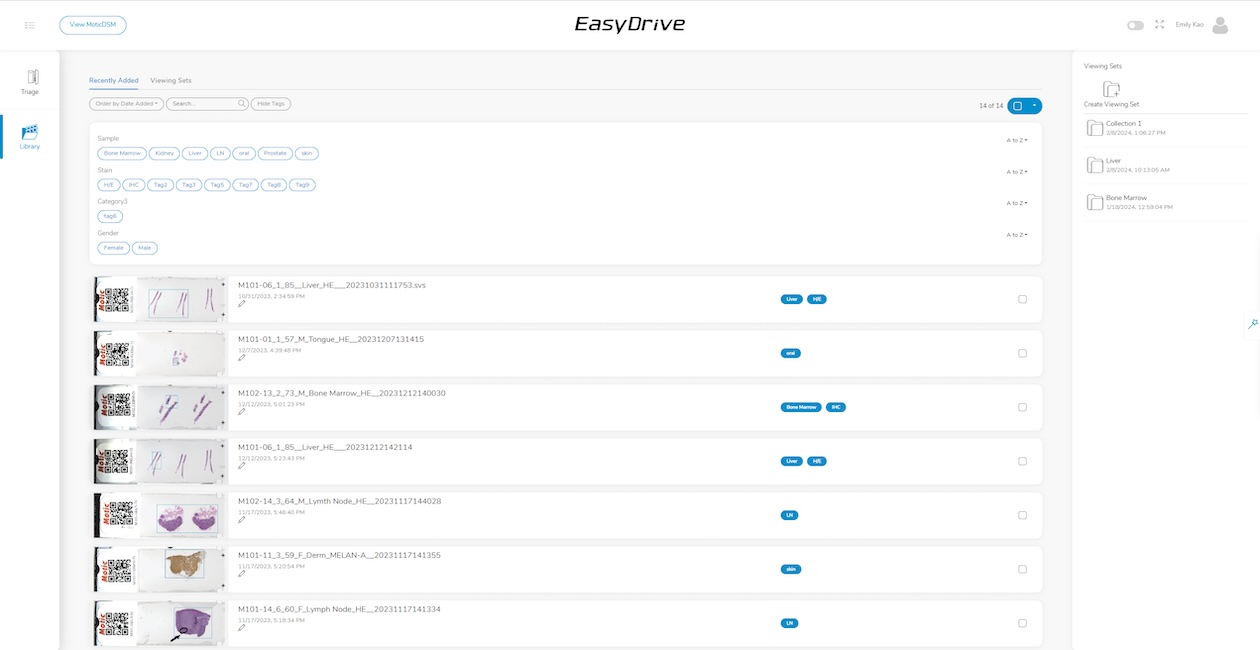
EasyDrive: Organizational Hub for Centralized Data Storage
EasyDrive seamlessly integrates with MoticEasyScan to enhance digital pathology workflows, offering a platform for sharing and managing data. It serves as an organized encyclopedia of digital pathology knowledge, enabling users to curate scanning runs systematically for easy access and sharing. With features like triage, users can effortlessly sort finished projects and isolate unneeded slides, while the library functionality allows for simple organization, search, and retrieval of slides. User can share slides via tiny URL-style link and archive unique samples for future study, ensuring that valuable data is preserved and accessible whenever needed. With MoticEasyScan and EasyDrive integration, laboratories can streamline their workflow, improve organization, and enhance collaboration in digital pathology.

LIS Connect: Customized Integration for Seamless Pathology Connectivity
LIS Connect provides Application Programming Interface (API) and remote viewing tools equipped with barcode parsing capabilities. It is tailored to provide efficient project planning and execution tools, along with scalability to meet requirements from entry-level to enterprise-level needs. With its intuitive user-friendly viewer integration and modern web viewer for pathology, LIS Connect seamlessly enhances your existing Lab Information System (LIS) with digital pathology functionality. By integrating the viewer with the LIS, it enables a smooth connection between electronic patient record-keeping systems and digital pathology software solutions. Additionally, LIS Connect can be optimized with barcode tracking to automate LIS data field population, thereby reducing manual entry errors and enhancing data accessibility and workflow efficiency.
FAQs
Yes, you may freely move between slides within the current tray and proceed to the next tray. However, you cannot navigate back to a previous tray once you have moved forward.
You can select from a range of open-source image file formats including .SVS, .JPEG, .TIFF, .DICOM, or .MDS.
Scan duratoin vary based on tissue size and objective. for instance, a 15x15mm scan at 40x magnification typically takes 100 seconds.
To accommodate increased demand, simply contact our support team to discuss upgrading your capacity from 24 slides to 100 slides.
Struggling with Your Workflow? Let’s Find a Solution.
Get a Free Consultation—Tailored to Your Needs.

Michael Kasten,
Motic Digital Pathology Expert
I am ready to provide my best support. Ask your question via the contact form or get in touch with me directly.
If you need assistance with your device, please contact Motic's
customer support
Is inefficiency slowing down your lab? Need a better way to manage your slides?
Connect with our experts for a personalized consultation and discover how MoticEasyScan Two can streamline your workflow, improve collaboration, and enhance efficiency.
Get answers within 24 hours and take the first step toward a smarter solution.

