USCAP 2026: Connected Digital Pathology Workflows for TMA and Real-Time Collaboration
Come and see Motic at booth #712 in San Antonio, Texas for the Annual USCAP 2026 meeting.
Improving interoperability in digital HER2 FISH enumeration a pilot evaluation
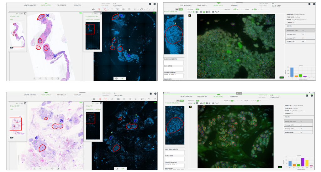
Tissue matching between H&E or IHC slides and HER2 FISH specimens allow for specific targeting of tumor regions with the highest protein expression, optimizing the high-magnification scanning of the FISH slide. Traditionally, this workflow was performed on the same scanning platform in the FISH lab. The present evaluation aims to assess the feasibility of matching a brightfield image acquired on one scanning platform, with a FISH image acquired on a different system, thus enabling the use of the region of interest marked by the pathologist on the H&E or IHC image without requiring a second rescan.
Combined manual reading and computer-aided quantitative analysis for the standardization of HER2 IHC scoring
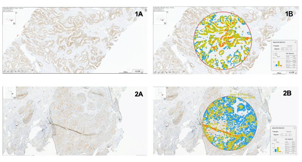
Computer-aided image analyses, when seamlessly integrated into the digital pathology workflow, are gaining in adoption. These methods, combined with manual reading, help standardize the reporting of IHC specimens, especially in cases where the tumor exhibits variable expression intensity. The evaluation aims to assess the usefulness of computer-aided quantitative analysis integrated into the laboratory workflow, focusing on its role as a second opinion for standardizing HER2 IHC scoring in breast cancer cases.
USCAP 2024 – United States and Canadian Academy of Pathology

Come and see Motic at booth #748 in Baltimore, M.D. for the 2024 USCAP (United States and Canadian Academy of Pathology).
Motic Digital Pathology and Applied Spectral Imaging (ASI) Announce First Projects in Joint-Venture to Create Integrated Digital Pathology Workflows for IHC and FISH Imaging through Validation with CorePath Laboratories
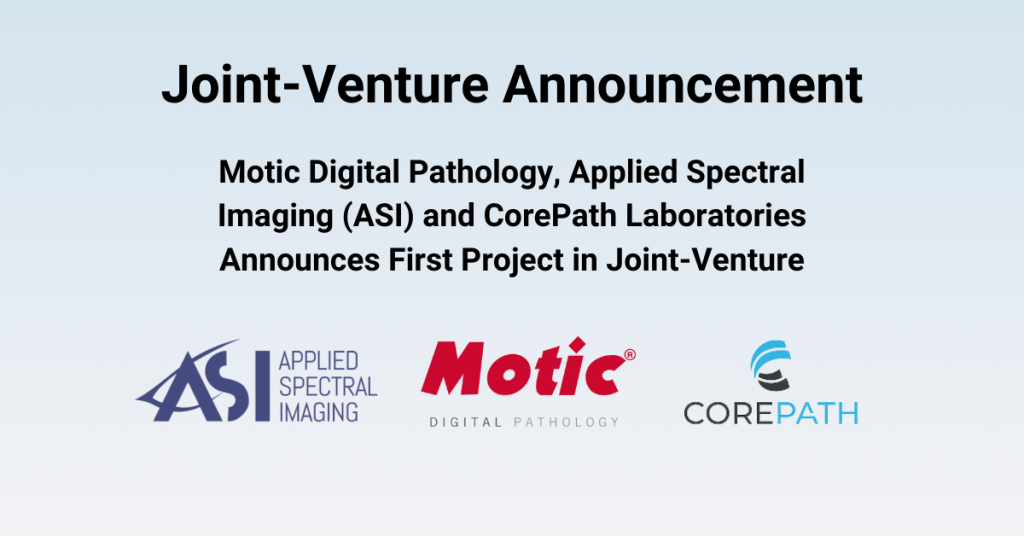
Motic Digital Pathology has selected ASI as its Joint-Venture partner for integrating its Whole Slide Imaging (WSI) seamlessly through ASI’s HiPath ProÔ and PathFusionÔ platforms for the analysis of brightfield and fluorescent images.
SFN 2023 – Society for Neuroscience
Come and see Motic at booth #1130 in Washington, D.C. for the 2023 Neuroscience Annual Meeting (SfN).
SITC 2023 – Society for Immunotherapy of Cancer
Come and see Motic at booth #329 in San Diego, C.A. for the SITC 2023 – Society for Immunotherapy of Cancer.