Combined manual reading and computer-aided quantitative analysis for the standardization of HER2 IHC scoring

Background and Introduction:
Computer-aided image analyses, when seamlessly integrated into the digital pathology workflow, are gaining in adoption. These methods, combined with manual reading, help standardize the reporting of IHC specimens, especially in cases where the tumor exhibits variable expression intensity. The evaluation aims to assess the usefulness of computer-aided quantitative analysis integrated into the laboratory workflow, focusing on its role as a second opinion for standardizing HER2 IHC scoring in breast cancer cases.
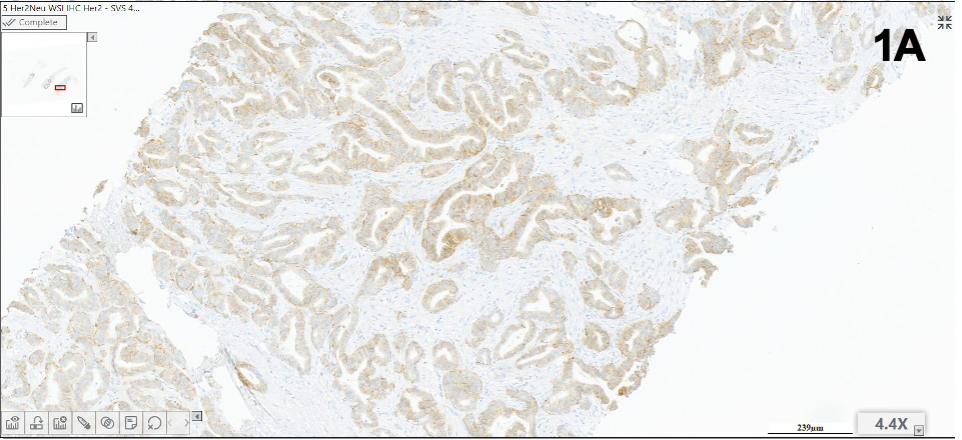
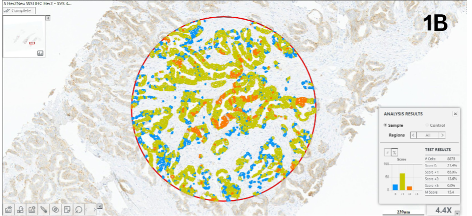
Figure 1: Representative examples of concordant HER2 case #5 (1) and discordant HER case #8 (2) featuring IHC staining (A) and computer-aided analysis (B)
Design and Method:
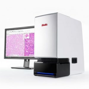
Five slides prepared for each breast cancer core biopsy specimen were stained with H&E, ER, PR, Ki67, and HER2. All slides were scanned with MoticEasyScan Infinity at 40X (0.26 um/px) and saved in SVS format. Certified pathologists examined the slides using conventional microscopy and digital imaging. HER2 FISH was requested to complete the evaluation of equivocal HER2 IHC cases. HER2 IHC images were further analyzed using the HiPath Pro scanner-agnostic software (Applied Spectral Imaging). Regions of interest marked on the H&E images were automatically transferred to HER2 specimens following tissue matching. Cells automatically identified as tumor cells were segmented and classified using a color-coded overlay. Computerized results were compared to manual readings, and in case of discrepancy, a second manual reading was performed.
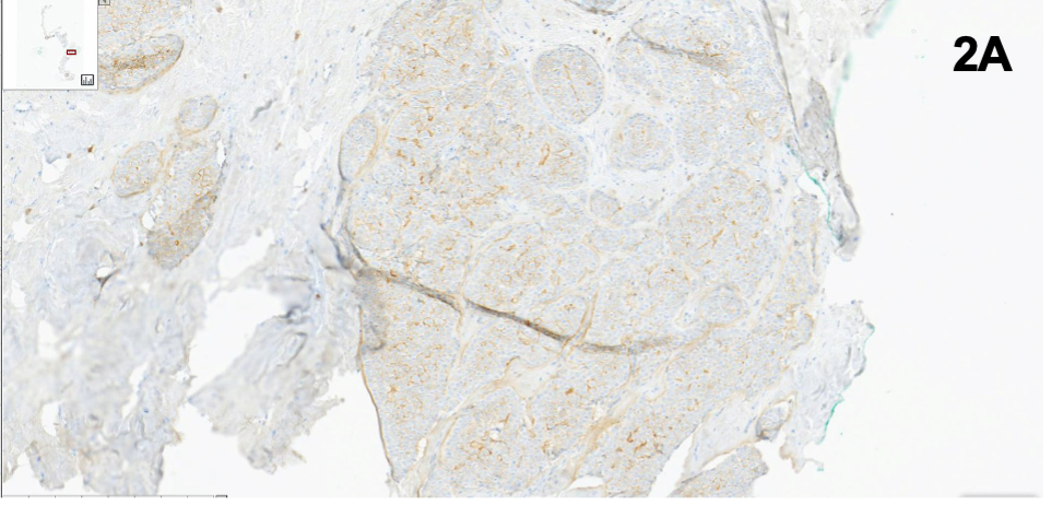
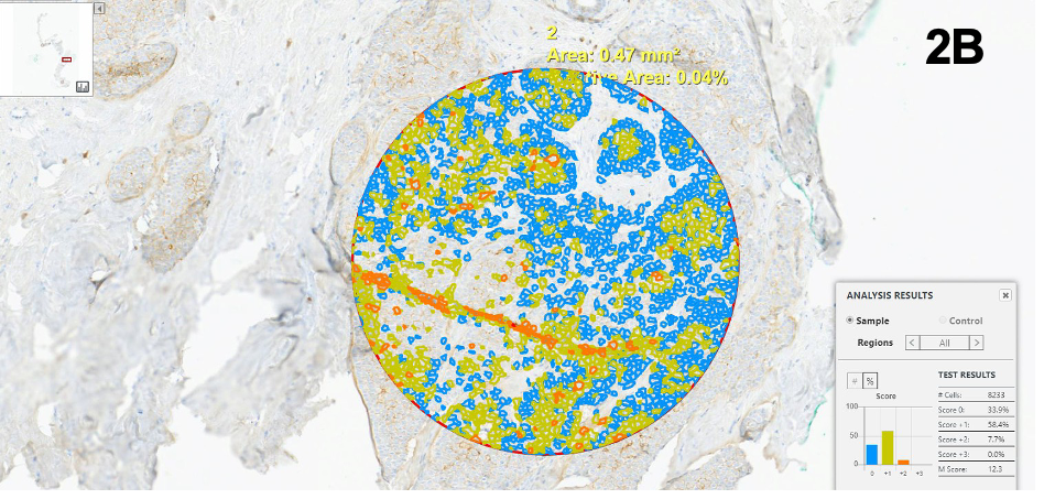
Figure 2: Representative examples of concordant HER2 case #5 (1) and discordant HER case #8 (2) featuring IHC staining (A) and computer-aided analysis (B)
Case # | ER | PR | Ki67 | HER2 Read 1 | HER2 Aided | HER2 Read 2 | HER2 FISH |
|---|---|---|---|---|---|---|---|
1 | + | + | <14% | 2+ | 2+ | NEG | |
2 | – | – | ≥14% | 0 | 0 | ||
3 | + | + | <14% | 0 | 1+ | 1+ | |
4 | + | + | ≥14% | 2+ | 2+ | NEG | |
5 | + | + | ≥14% | 2+ | 2+ | NEG | |
6 | + | – | ≥14% | 3+ | 3+ | ||
7 | + | + | ≥14% | 0 | 0 | ||
8 | + | + | ≥14% | 2+ | 1+ | 2+ | NEG |
9 | + | + | ≥14% | 3+ | No focus | ||
10 | + | + | ≥14% | 2+ | 3+ | 3+ | POS |
11 | – | – | ≥14% | 0 | 1+ | 1+ | |
12 | + | + | ≥14% | 1+ | 1+ | ||
13 | + | + | <14% | 0 | 0 | ||
14 | – | – | ≥14% | 0 | 0 | ||
15 | + | + | ≥14% | 1+ | 0 | 1+ | |
16 | + | + | ≥14% | 3+ | 2+ | 3+ | |
17 | – | + | ≥14% | 2+ | 2+ | POS | |
18 | – | – | ≥14% | 2+ | 2+ | POS | |
19 | + | + | ≥14% | 0 | 0 | ||
20 | – | – | ≥14% | 3+ | 3+ |
Table 3: IHC profile of study cases including manual reading of ER, PR, Ki67 and HER2 (reading 1 and repeated reading 2 for discrepant cases), as well as computer-aided HER2 IHC scoring. HER2 FISH results are provided for all equivocal cases.
Results:
Twenty biopsy specimens from 20 patients were included in this evaluation. Nineteen samples had a diagnosis of invasive ductal or lobular carcinoma, and one of metaplastic carcinoma with chondroid differentiation. The ER, PR, Ki67, and HER2 IHC profiles of the cases are detailed in Table 1. As shown in this table, the first manual reading of HER2 IHC diagnosed 4 cases as positive (3+), 9 as negative (0 or 1+), and 7 as equivocal (2+). FISH was performed on all equivocal cases, confirming 3 as HER2 positive and 4 as HER2 negative.
Computer-aided HER2 scoring was obtained for 19 out of 20 images, as one image out of focus was removed from the analysis. Concordance with the first manual reading was observed in 13 cases. During the second manual reading following computerized analysis, two cases scored as HER2 (0) were re-scored as HER2 (1+), matching the software assessment. A third case diagnosed as equivocal (2+) was re-scored as (3+) following the review of the computerized analysis. This case was later confirmed as FISH positive. The diagnosis of the remaining 3 cases remained unchanged after the review of computerized results.
Conclusion:
This evaluation demonstrates the potential usefulness of computer-aided scoring as a second opinion in reporting HER2 IHC in breast cancer, particularly in cases of low HER2 expression. Furthermore, it illustrates how the combined use of manual reading and computerized analysis may help standardize IHC assessment and potentially reduce inter-observer variability.