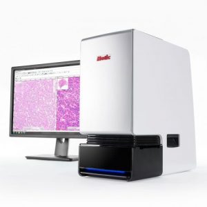
Digital pathology is becoming an increasingly prevalent tool in laboratories and hospitals beyond the early pioneers in the West. The ability to receive rapid, real-time support from a wide variety of specialized pathologists has strengthened the diagnostic confidence of many physicians, resulting in better patient outcomes and enhanced quality care. And as a training tool, Whole Slide Images have allowed mentors and trainees to seamlessly access a wide range of digitized tissue samples, eliminating the need to laboriously sift through conventional glass slides. In this blog, we are shining the spotlight on an organization that recently adopted this technology: Obafemi Awolowo University (OAU) in Nigeria. Motic, in partnership with the American Society for Clinical Pathology, deployed a MoticEasyScan Pro 6 scanner to OAU for remote consultation use. This project was graciously funded by Memorial Sloan Kettering Cancer Center (MSKCC) to support cancer patients and advance cancer research in Nigeria.
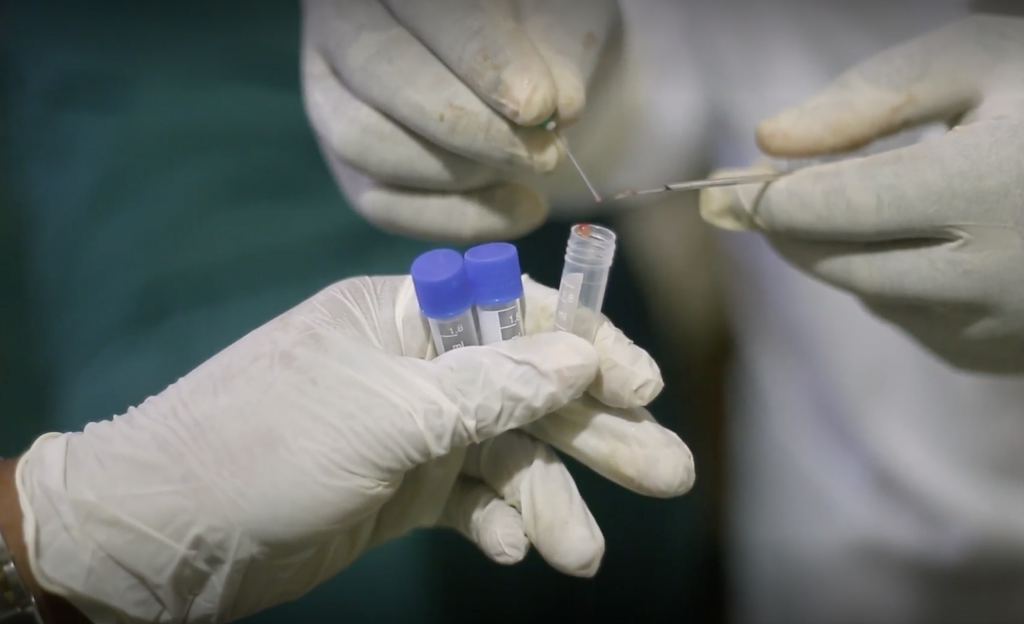
Samples are taken and sent to the lab to be prepared
Strengthening Diagnostic Confidence
Prior to the implementation of digital pathology, Dr. Olusegun Isaac Altaise, Professor of Surgical Oncology and Gastroenterology at OAU, said immunohistochemistries (IHC) were performed locally by the histopathologists at the clinic. For validation purposes, unstained slides were shipped to MSKCC for IHC comparison. With traditional microscopy, however, receiving secondary opinions is limited to only a handful of pathologists, which can undermine confidence in the diagnostic results.
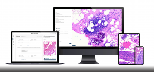
Thanks to whole-slide imaging and MoticFlow—the slide-sharing software suite, Dr. Altaise can now scan every case slide onsite at OAU and digitally distribute these cases to doctors anywhere from Nigeria to New York for instant secondary diagnostic support.
Ultimately, the most significant benefits of this new program to Nigerian pathology is a major leap in confidence that Dr. Altaise has in his department’s signouts, the ability to receive real-time support from pathologists in the West, and a drastic improvement in the health outcomes and quality of care for his patients.
“The treatment of our cancer patients can never be better than what we have now, because not only do we know what we’re treating, but we have support,” expresses Dr. Altaise.
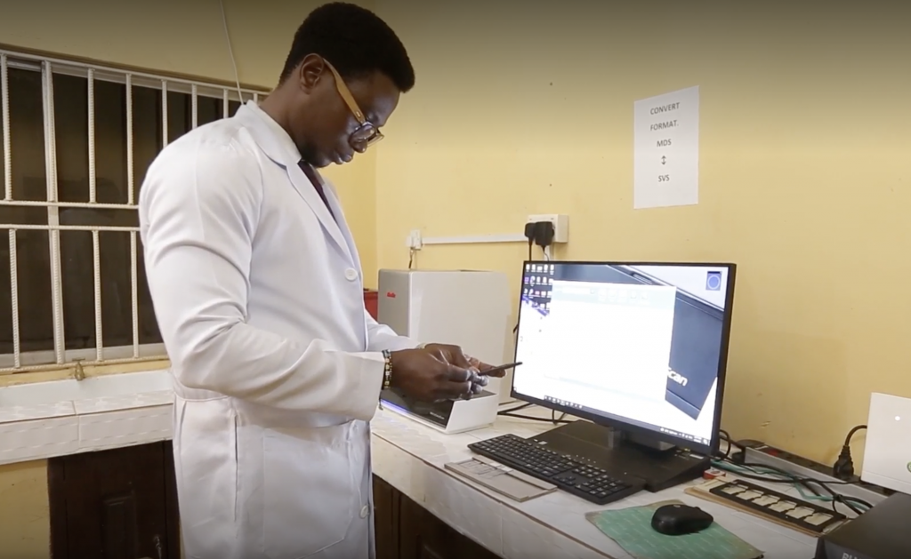
Slides are loaded into the scanner for digitization
Motic in Nigeria
The images generated from the MoticEasyScan Pro 6 scanner have helped in all spaces of their practice, said Dr. Adeyemi Abiola Adefidipe, Consultant Histopathologist at OAU. One ancillary benefit is that, because the images of every slide made at the lab are stored digitally, doctors can provide timely feedback to lab personnel, even from geographically remote locations, and identify areas where artifacts are being formed by slide production errors. According to Oyasola Adedayo O., Chief Medical Laboratory Scientist at OAU, this direct feedback loop has greatly improved output quality at the laboratory, and his team are very grateful for the scanner as a result.
With the advent of digital pathology, there is increased opportunity for collaboration between pathologists and labs from virtually anywhere in the world. This means streamlined laboratory operations, reduced treatment errors, and the delivery of accurate diagnostic results with rapid turnaround times. And of course, the sooner a diagnosis is made, the sooner life-saving treatment can be administered to patients.
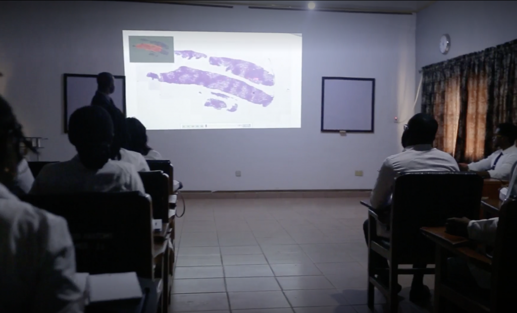
Breast Cancer Echo and Colorectal Echo taught using digitized slides
Transforming Medical Education in Nigeria
Digital pathology not only provides assistance with primary diagnoses and secondary opinions, but also with medical training. OAU has two training programs: Colorectal Cancer Echo and Breast Cancer Echo. More than 50-100 professionals from all throughout Nigeria can participate remotely in these training programs, allowing the university to hold more frequent group seminars and welcome a much larger cohort of medical residents. By facilitating remote training, digital pathology expands access to medical education in remote, underserved communities.
Furthermore, the use of digital pathology allows for effortless searching through a Motic’s digitized library, compared to manually sorting glass slides in a physical archive. This feature allows Dr. Altaise to save and store case slides within the scanner’s supporting software, granting pathologists and residents at OAU quick and easy access to the entire catalog of the hospital’s slides. It now takes minutes, rather than hours, to put together review sets for tumor boards, presentations, and even slides for one-on-one discussions. With a myriad of case slides at their disposal, medical residents can now properly highlight and study the most challenging cases quickly, smoothly, and conveniently.
There are now more ways than ever before to review cases in pathology training thanks to the educational possibilities made accessible by digital pathology. Digital pathology is optimizing Nigerian medical education, serving as an invaluable instructional tool for medical residents learning how to diagnose cancer.
Long considered a luxury medical device reserved for affluent nations, digital pathology is becoming a practical reality in Nigeria, redesigning medical education and aiding in the reformation of the country’s cancer treatment initiatives.

