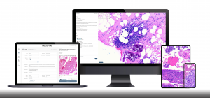
At the start of this pandemic, we highlighted how different digital pathology tools can enable remote work and collaboration. We’d now like to delve into how superior autofocus plays a crucial role in live remote consultations, especially for time-sensitive procedures such as Frozen Sections, Fine-Needle Aspirations (FNAs), and ROSE.
Using an analog microscope
Every pathologist is familiar with this:
When using an analog microscope, one hand operates the stage controls or taps the slide for movement; the other hand controls focus. Both hands work in concert to keep the sample in focus as you move, or, in the case of cytology, the focusing is also used to scan through thick samples to locate cells or parasites suspended in the slide medium.
Every microscopist has been trained this way for the past two hundred years, but how does the handling change when you go digital?
How does a machine handle focusing?
When manipulating a slide digitally, the user controls the x- and y-axes to move the sample, while the z-axis is reserved for focusing. Most systems today have integrated autofocusing for ease of use. Typically, these focusing protocols operate on only one criteria: whether the image is in or out of focus.
If the image is in focus, everything is fine. If the image is out of focus, however, the camera tries to refocus by sampling several different z-levels and picks the best one to display. We all know this feature, it’s in our DSLRs, smartphone cameras, and in every scanning system for digital pathology. And it works well!
… Except for time-sensitive procedures, where a massive problem arises: at high magnifications, even the slightest variations in sample thickness can take the image out of focus. Not only does this require frequent refocusing on part of the camera, the depth of field at such magnifications is only around 1 um, making it very time consuming to find the correct plane (pathology samples can vary between 5-100 um in thickness). Additionally, these Frozen Section or FNA samples are often not flat, so in the course of evaluating a single slide, a machine may need to refocus the sample up to 30-40 times.
That’s almost 1-2 minutes lost per slide waiting for autofocus to do its work.
One workaround is to turn off autofocus and attempt to control the sample yourself. However, unlike a microscope, remote digital systems have to contend with network lag, which makes real-time adjustment a frustrating and inefficient experience. The other solution is to have a trained technician control the microscope for the pathologist, who views the live video stream, but has no control over the sample.
Neither option is truly viable as a long-term solution, and this is the primary reason why adoption of remote access systems is not yet mainstream.
A superior autofocus that just works
With these issues in mind, our mission was clear: build a superior live remote viewing system that uses a new type of autofocus, one that keep the image in focus instantaneously, even for uneven samples.
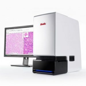
The engineers at Motic answered with a one-of-a-kind continuous autofocus system, which replaces the traditional, clunky autofocus with a smarter design using multiple cameras and next-gen algorithms. No matter how the sample is moved, the focus continually adapts to the contours of the sample in real-time, keeping the view perfectly in focus wherever you go.
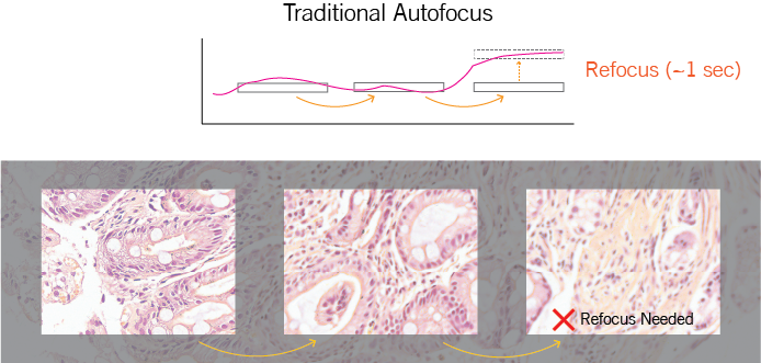
With traditional autofocus, movement across an uneven sample quickly results in a view that’s out of focus.
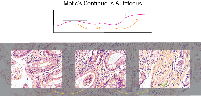
Compared to traditional autofocus systems, Motic’s refocuses in one-step.
With this revolutionary feature at the core of Motic’s FS-Live telepathology system, pathologists are empowered to review case slides interruption-free, and focus solely on analyzing and performing the readout. By making remote reviews as intuitive as operating an analog microscope, we hope to help pathologists fully realize the benefits of live digital consultations.
Dr. Enric Solans, who is a practicing general pathologist in Chicago, speaks to the challenges that FS-Live has helped him overcome:
“Before going digital, I would constantly have to manually adjust the focus of my microscope while navigating through the slide and changing magnifications… I would briefly lose clear view of the tissue which was a bit frustrating.
Motic’s viewing software allows me to view the sample clearly, even with folds in the tissue or wet slides. I no longer have to re-adjust, as the slide is automatically in focus the entire time, saving me time on consultations.”
Having an autofocus for live slide viewing that “just works” can greatly ease the transition from analog consultations to a digitized, remote workflow. We firmly believe the best way to evaluate the speed and effectiveness of any telepathology system is to experience it for yourself.
Book a virtual demo with us, and see first-hand how our autofocus can facilitate effective, timely remote procedures at your institution!