Improving interoperability in digital HER2 FISH enumeration a pilot evaluation
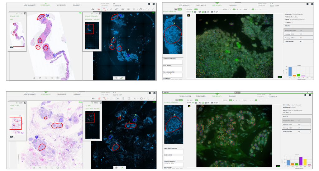
Tissue matching between H&E or IHC slides and HER2 FISH specimens allow for specific targeting of tumor regions with the highest protein expression, optimizing the high-magnification scanning of the FISH slide. Traditionally, this workflow was performed on the same scanning platform in the FISH lab. The present evaluation aims to assess the feasibility of matching a brightfield image acquired on one scanning platform, with a FISH image acquired on a different system, thus enabling the use of the region of interest marked by the pathologist on the H&E or IHC image without requiring a second rescan.
Combined manual reading and computer-aided quantitative analysis for the standardization of HER2 IHC scoring
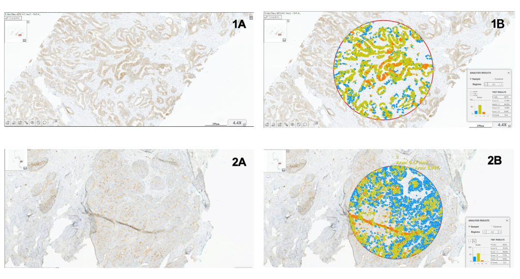
Computer-aided image analyses, when seamlessly integrated into the digital pathology workflow, are gaining in adoption. These methods, combined with manual reading, help standardize the reporting of IHC specimens, especially in cases where the tumor exhibits variable expression intensity. The evaluation aims to assess the usefulness of computer-aided quantitative analysis integrated into the laboratory workflow, focusing on its role as a second opinion for standardizing HER2 IHC scoring in breast cancer cases.
USCAP 2024 – United States and Canadian Academy of Pathology

Come and see Motic at booth #748 in Baltimore, M.D. for the 2024 USCAP (United States and Canadian Academy of Pathology).
SFN 2023 – Society for Neuroscience
Come and see Motic at booth #1130 in Washington, D.C. for the 2023 Neuroscience Annual Meeting (SfN).
SITC 2023 – Society for Immunotherapy of Cancer
Come and see Motic at booth #329 in San Diego, C.A. for the SITC 2023 – Society for Immunotherapy of Cancer.
Motic in Haiti: The Benefits of Telepathology in Underserved Communities

By introducing affordable telepathology platforms, Motic is revolutionizing healthcare in underserved communities like Haiti. The integration of digital pathology systems at HUM has addressed the lack of pathologists and significantly improved the efficiency and accessibility of cancer treatment. Motic’s commitment to making healthcare accessible in underserved regions demonstrates the potential of technology to bridge gaps in medical services and improve outcomes for patients in need.
WSI & Digital Pathology Lab Equipment: A Comprehensive Guide
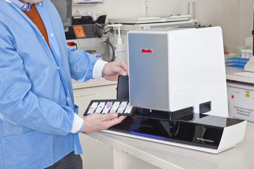
Digital pathology refers to the use of technology to digitally capture, store, and analyze tissue samples in a laboratory setting. This technology has revolutionized the field of pathology and offers many benefits over traditional methods, including increased speed, accuracy, and efficiency. Pathologists no longer need to spend hours hunched over a microscope or spend time […]
5 Scanners for $100k: How to Utilize 5 Scanners in 5 Different Applications
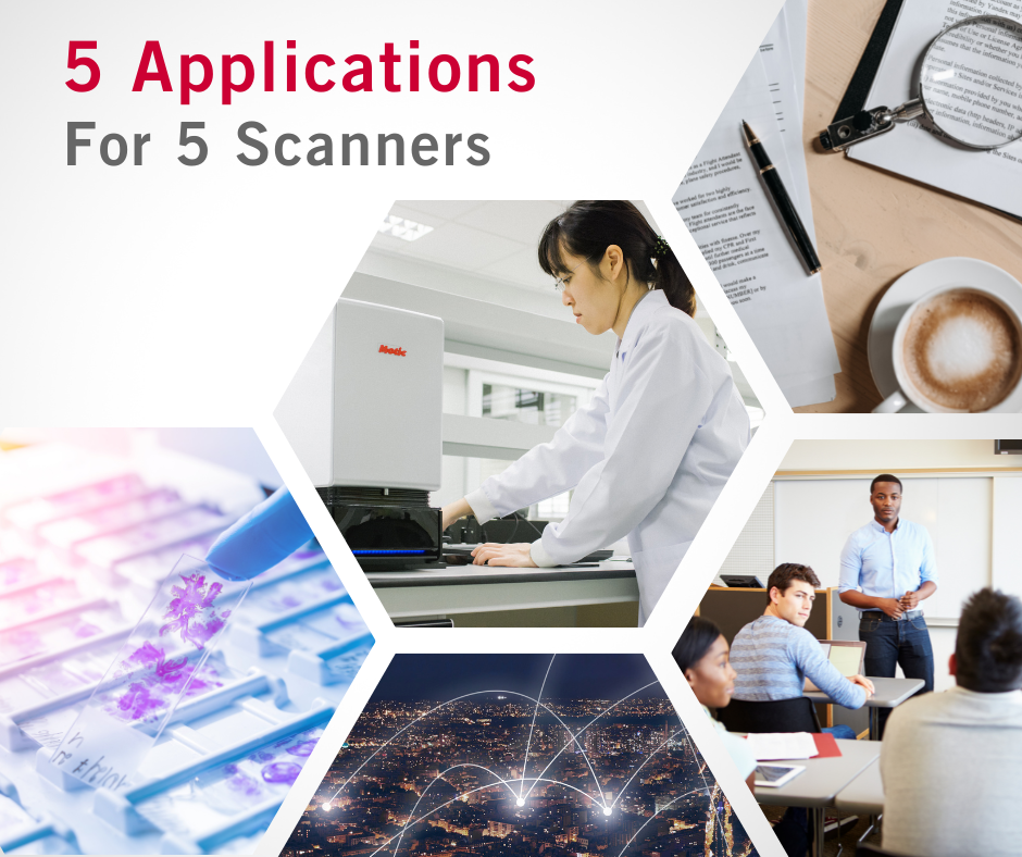
In honor of our fifth anniversary and our ongoing commitment to democratizing access to digital pathology worldwide, we will be offering the biggest promotion in our company’s history
Come Celebrate 5 Years of MoticEasyScan with Our Special Anniversary Offer!
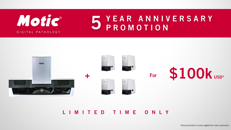
Motic Digital Pathology will be celebrating its 5 years of revolutionary digital pathology USCAP 2023 Motic began our digital pathology journey 5 years ago at USCAP 2018, when we announced a trio of novel scanners specifically designed for first adopters: MoticEasyScan One, MoticEasyScan Pro 6, and MoticEasyScan Infinity. Hundreds of pathology labs have since been […]
The 112th Annual Meeting of the Japanese Society of Pathology

April 13 to 15, 2023
Japani
Shimonoseki City Lifelong Learning Plaza (DREAM SHIP)