Telecytology has historically lagged behind histology applications as digital pathology grew toward widespread acceptance in the last decade. While recent technological advancements are rapidly closing the gap in affordability and ease-of-use for telecytology, many cytopathologists still wonder if the digital pathology revolution includes them. Luckily, the answer today is a resounding yes.
Telecytology, Then and Now
To understand why digital histology took off before cytology did is really to understand a tale of two file sizes.
In the early days of digital pathology, hardware companies built scanners with the flat, even slides used by histopathologists in mind. Flat tissue was easier to scan since the necessary information is typically already found on one focal plane. The image files, while large, were not prohibitively so for sharing and storage.
In contrast, the cytopathology workflow was — and still is — more difficult to capture digitally. Cytologists need to see multiple layers of a cell to render judgments, so generating single-layer, two-dimensional images is ineffective for their use. Scanner makers have responded by creating z-stacking features, which can focus through multiple layers. However, z-stack scans contain far more digital information than flat slides do, meaning the cost of data storage quickly becomes a concern. Uploading and downloading such large files for consultation also takes more time, though it’s still an improvement on mailing glass slides!
The Growing Need for Telecytology
Enter telecytology. With the increased use of ROSE and FNA procedures, the overall cytology caseload has grown. While these intraoperative assessments are not as time-consuming as rendering diagnoses, they are time-sensitive and nevertheless represent demands on the cytopathologist’s overall workload. (A distinction that the College of American Pathologists recognized in a creative 2018 rules change that sets adequacy screening limits based on time rather than the number of slides assessed.)
While the ideal scenario is always to have the cytopathologist onsite at the surgical suite to perform the adequacy reading, this in-person assessment is not always practical, especially for larger hospital networks or labs with limited cytopathology personnel.
Laboratories have responded in creative ways, proving that necessity is the mother of invention. Early versions of telecytology consisted of live-streaming slide images to the cytopathologist via cameras either built into or mounted on microscopes. Technicians, directed by cytopathologists over the phone, control the slide, panning around the field of view and focusing through different layers as needed.
The Advantages of Telecytology
While a technician-cytopathologist workflow has worked quite well for enabling telecytology, designing true telecytology solutions that allow pathologists to control the slide image from afar is a commonsense next step in digital pathology. For one, it is not only the cytopathologist’s time that gets tied up with ROSE and FNA. The cytotechnician must also interrupt their work to prepare slides, connect with the offsite pathologist, and control the microscope.
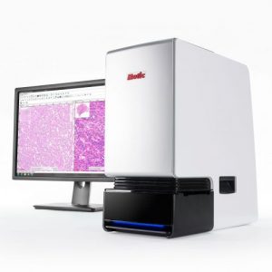
Real-time remote telecytology applications, like Motic’s FS-Live Telepathology System, save time for cytotechnicians. With this live workflow, techs still prepare the slide for reading. But once they’ve loaded the slide into the scanner, the remote cytologist takes over the controls, using the scanner as a robotic microscope from anywhere. It’s a win-win for both individuals. The cytopathologist saves time by not having to direct the technician. Meanwhile, the technician can move on to other tasks, including prepping more slides.
No Longer Left Out: Designing Telecytology Solutions That Cytologists Love
When it comes to telecytology, scanner companies have the opportunity not to repeat the mistake of building histology solutions that they then try to back-engineer for cytology. In other words, we must design telecytology solutions for cytologists first.
This concept seems like it should be simple, but in our conversations with cytopathologists, we’ve found that it’s often far from obvious. Cytology specialists still tend to assume that digital pathology companies have nothing to offer them. And no wonder, when cytology has commonly been treated as an afterthought.
When our engineering team designed FS-Live, they prioritized the following features that would make it easy for cytopathologists to use the system remotely for either adequacy or consultation:
- Continuous autofocus with manual override: Cytologists can use the always-in-focus image for a quick scan and then step through various layers with their mouse scroll wheel.
- Stain compensation: Is the sample over-or under-stained? That’s not a problem with our gamma compensation feature, which allows the user to digitally lighten or darken the image to see cell morphology and nuclear details.
- Z-stack snapshot: Isolate an area of interest and generate a file with all cell layers in just a few seconds. The user simply sets the number of focus layers, the step increments, and then hits the spacebar to produce a quick z-stack image without lengthy scanning time, enormous file sizes, or needing to leave the live-view interface.
- 4X objective option: Offers a large initial field of view without compromising image quality at higher magnifications.
- Drop markers: Technicians can still note areas of interest before the cytopathologist takes over.
There are few experiences more gratifying to a technology company than opening a door for an entire set of users who’d previously been left behind by exciting innovations. In the case of digital pathology, solutions for easy, efficient, and affordable telecytology are finally here.
Do you want to bring telecytology to your lab with Motic’s FS-Live System? Contact us today for a free demo on all our features built specifically for cytopathology!