The field of pathology is undergoing a transformative shift with the emergence of remote frozen sections (RFS), a groundbreaking concept enabled by digital pathology. Frozen section analysis has played a crucial role in intraoperative diagnosis, but it traditionally required on-site pathologists. By requiring pathologists on-site, there is an increase in travel time which could lead to potential delays and limited access to expert opinions.
However, remote frozen section leverages digital pathology to overcome these challenges, by enabling pathologists to analyze tissue samples and provide real-time diagnoses from anywhere in the world. This paradigm shift has the potential to revolutionize this industry, offering benefits such as enhanced access to expertise, reduced turnaround times, and improved collaboration among pathologists.
Traditional Frozen Section Procedure
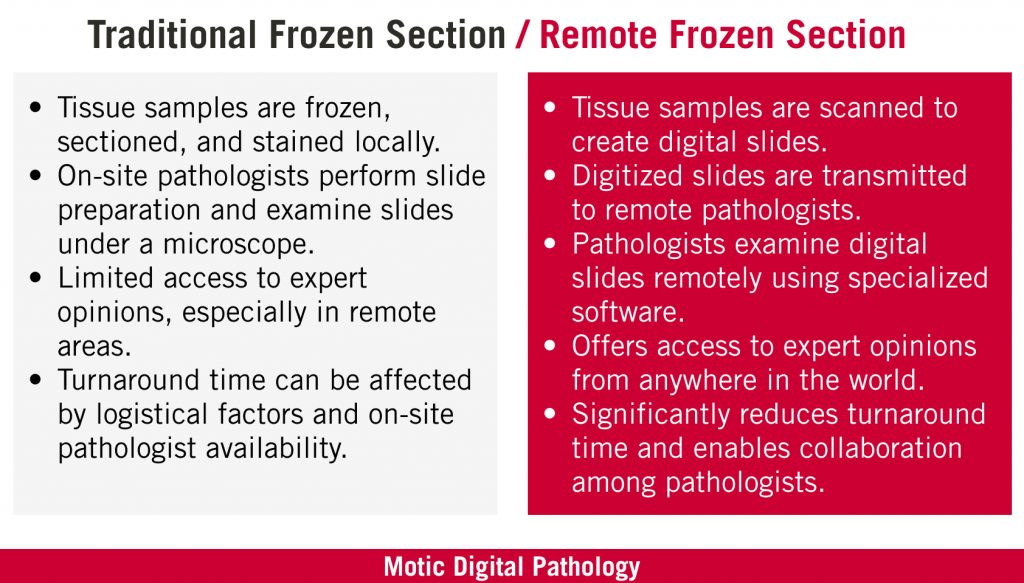
The traditional frozen section process (FSP) has long been a cornerstone of intraoperative diagnosis in pathology. It involves a sequential workflow that includes freezing, sectioning, staining, and microscopic examination of the tissue.
The process begins with the rapid freezing of fresh tissue specimens using a cryostat, which allows the tissue to be hardened and prepared for sectioning. Thin slices, or sections, of the frozen tissue are then cut using a microtome and mounted onto glass slides. These slides are typically stained using various dyes to enhance the visualization of cellular structures and tissue morphology. Once stained, the slides are examined under a microscope by an on-site pathologist, who analyzes the cellular features and makes a real-time diagnosis.
Challenges & Limitations
While the traditional frozen section process has been valuable in providing intraoperative diagnostic information, it is not without its challenges and limitations. Time constraints pose a significant challenge, as the process requires the immediate preparation and examination of tissue samples during surgery. This urgency can place considerable pressure on pathologists to deliver rapid diagnoses, potentially affecting the accuracy and thoroughness of their evaluations.
The traditional method necessitates the physical presence of on-site pathologists in the operating room or nearby pathology laboratory. This requirement poses logistical challenges, especially in rural or underserved areas where access to specialized pathology services may be limited. In such cases, the lack of immediate access to expert opinions can lead to delays in obtaining critical diagnostic information, potentially impacting surgical decision-making and patient outcomes.
Also, the reliance on on-site pathologists for frozen section analysis can strain resources and increase the burden on pathology departments. The need for dedicated personnel to be available at all times, including nights and weekends, to perform frozen sections can be challenging to sustain, particularly for smaller healthcare facilities with limited staffing capacities.
These challenges underscore the need for innovative solutions that can address the limitations of the traditional frozen section process. Remote frozen section, enabled by digital pathology, has emerged as a promising approach to overcome these hurdles. It offers improved access to expert opinions, faster turnaround times, and enhanced collaboration among pathologists regardless of their physical location.
Remote Frozen Sections with a Digital Pathology Instrument
Remote Frozen Sections is a practice in pathology where pathologists analyze frozen tissue sections during surgical procedures, providing rapid patient information to guide surgical decision-making. This technique is particularly valuable in cases where there is an urgent need for immediate pathology assessment, such as determining the nature of a tumor or evaluating the margins of a resected specimen.
The advantages are clear:
- Remote frozen sections facilitate access to expert opinions regardless of geographical location, ensuring patients benefit from the insights of experienced pathologists, even in underserved areas.
- The rapid evaluation of digitized slides significantly reduces turnaround times, expediting surgical decision-making.
- Remote frozen section promotes collaboration and knowledge sharing among pathologists by breaking down geographical barriers, leading to enhanced diagnostic accuracy and fostering continuous learning within the pathology community.
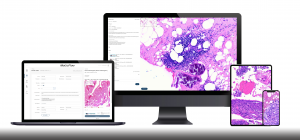
With the advent of WSI technology, pathologists can now remotely access and evaluate frozen tissue sections in real-time, enabling them to provide Remote On-Site Evaluation support from a remote location. Through telepathology, pathologists can view high-resolution digital slides of the frozen sections, allowing for detailed examination and interpretation.
Rapid On-Site Evaluation (ROSE)
ROSE is a technique used in pathology to provide quick diagnostic information during a medical procedure, such as a biopsies. WSI has enabled pathologists to perform ROSE remotely, using digital images instead of physical samples.
WSI allows for the transmission of high-quality images over vast distances, allowing pathologists to analyze slides with the utmost accuracy and efficiency regardless of their physical location. Therefore, the use of WSI is closely connected to ROSE, and has revolutionized the way that pathologists can conduct remote evaluations on tissue samples.
The implementation of WSI for ROSE in RFS also streamlines workflows and enhances efficiency. It eliminates the logistical challenges and delays associated with transporting physical slides, ensuring that the pathological assessment can be performed rapidly. This streamlined process not only saves time but also contributes to improved patient care by reducing the time patients spend under anesthesia during surgical procedures.
Tradition Frozen Section VS Remote Frozen Section
The main difference between the traditional frozen section and remote frozen section lies in the approach used for slide preparation, examination, and analysis. Here’s a breakdown of the key distinctions:
Slide Preparation
In the traditional frozen section approach, tissue samples are subjected to slide preparation within the pathology laboratory at the surgical site. On-site pathologists are responsible for the entire slide preparation process, which entails freezing the tissue, sectioning it into thin slices, and applying appropriate stains. This local slide preparation allows for immediate examination under a microscope by the pathologist present at the facility.
In contrast, the remote frozen section takes advantage of digital pathology advancements. While the tissue samples are still frozen, the slide preparation process takes a different path. Instead of local preparation, the frozen tissue samples are remotely viewed by a robotic microscope By eliminating the need for on-site slide preparation, the remote frozen section streamlines the process, enables efficient transmission of digital slide viewing, and opens the door for remote analysis by pathologists from any location.
Slide Examination and Diagnosis
In the traditional frozen section process, the pathologist conducts the examination of stained slides on-site, directly observing them under a microscope. This in-person evaluation allows for physical slide viewing, but it necessitates the pathologist’s physical presence at the location where the slides were prepared.
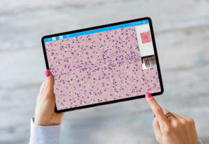
In contrast, remote frozen section in digital pathology revolutionizes the way slide examinations are performed. Pathologists have access to the digitized slides through a telepathology platform, which enables remote viewing and analysis. Utilizing specialized software on a computer or other viewing devices, pathologists can examine the digital slides from any location. This remote access empowers pathologists to view slides in real-time and eliminates the need for physical proximity to the site of slide preparation. The ability to analyze the images remotely enhances flexibility, collaboration, and access to expertise, revolutionizing the frozen sections process.
Access to Expert Opinions
In the traditional frozen section process, the availability of expert opinions is constrained to the expertise of the on-site pathologist conducting the frozen section. This limitation can be especially challenging in remote or underserved areas, where access to specialized pathology expertise may be scarce or non-existent.
On the other hand, the remote frozen section in digital pathology introduces a transformative change by providing unparalleled access to expert opinions. With the digitization of slides, it becomes effortless to share the digital images with pathologists globally. This facilitates remote consultation and collaboration among pathologists regardless of their geographical location. Remote frozen section opens doors to a broader pool of expertise, allowing pathologists to seek and provide consultations from a diverse range of specialists. The ability to tap into a wealth of knowledge and experience across different regions significantly enhances the quality and accuracy of slide analysis, regardless of geographical limitations.
Turn around Time and Efficiency
The traditional frozen section method’s turnaround time is influenced by several factors, including the time required for on-site slide preparation, logistical challenges related to slide transport, and the availability of a pathologist. Delays can have a direct impact on surgical decision-making and patient care, potentially leading to prolonged procedures or delays in initiating appropriate treatments.
In contrast, the remote frozen section in digital pathology brings about a remarkable improvement in turnaround time and efficiency. By digitizing slides, they can be rapidly transmitted to pathologists located remotely. These pathologists can then examine the digitized slides remotely using specialized software, enabling them to promptly analyze the slides. The streamlined process expedites a pathologist’s workflow, facilitating faster decision-making in the operating room. The reduction in turnaround time not only benefits surgical procedures but also enhances overall patient care by minimizing potential delays in treatment initiation and improving the efficiency of healthcare delivery.
While the traditional frozen section relies on on-site slide preparation and examination, remote frozen section in digital pathology eliminates the need for physical presence. It leverages digital slide scanning, remote viewing capabilities, and telepathology platforms to enable access to expertise from any location, reducing turnaround times and facilitating collaboration among pathologists.
Remote Frozen Section Workflow
Performing a remote frozen section using digital pathology involves a systematic and efficient workflow. Here is a step-by-step description of the process:
Tissue Sampling: The surgical team collects the tissue sample during the procedure, ensuring proper handling and preservation to maintain its integrity.
Freezing and Preparation: The tissue sample is promptly frozen using a cryostat or similar device, preserving its cellular structure. The frozen tissue block is then carefully prepared for sectioning.
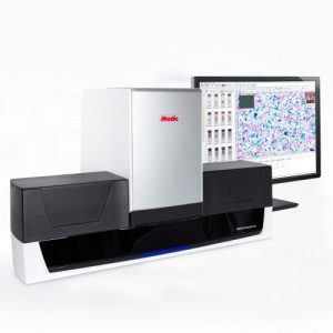
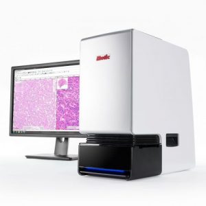
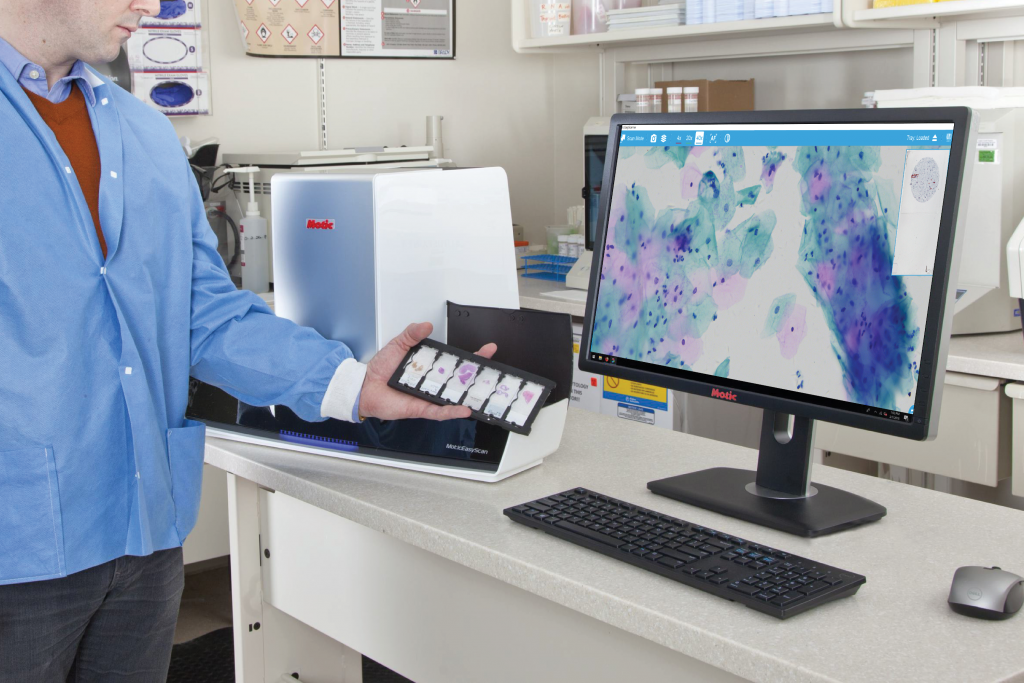
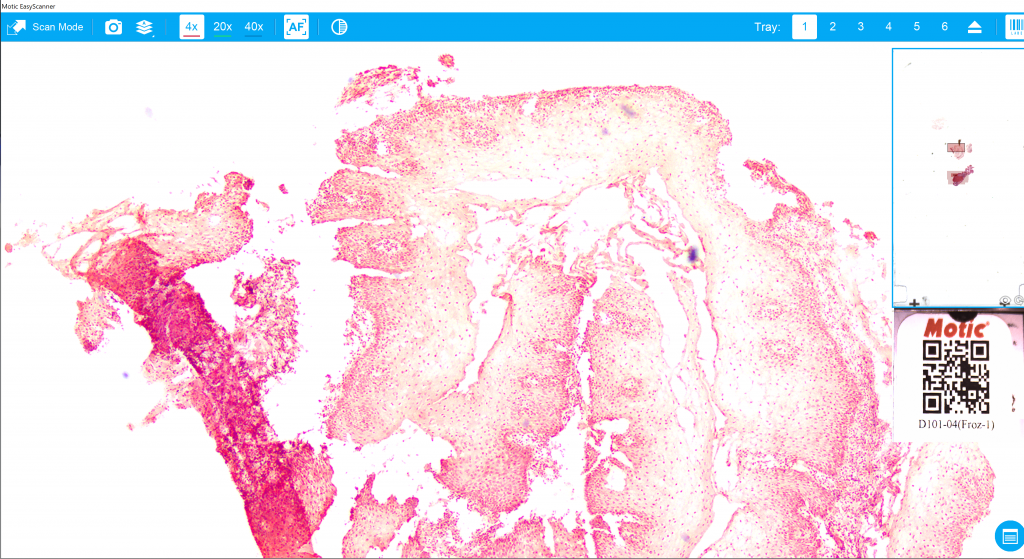
High-Resolution Slide Scanning: The prepared tissue sections are viewed under a digital pathology instrument with live viewing capabilities., for example, with MoticEasyScanPro6 slides can be viewed live remotely from anywhere anytime.
Remote Examination: Pathologists view the digitized slides through the telepathology platform using specialized software such as FS-Live. They can examine the slides remotely, zoom in on specific areas of interest, and analyze the cellular features to accurately analyze slides in real-time
Quality Assurance Measures: Quality assurance is crucial in remote frozen section. Pathologists ensure that the digital images accurately represent the original tissue slides and that the scanning process maintains high resolution and image fidelity. Regular calibration and quality control checks are performed to maintain the accuracy and consistency of the digital slide viewing.
Secure Communication Channels: To ensure patient privacy and confidentiality, secure communication channels and encrypted data transmission protocols are employed for the exchange of patient information and results.
Compliance with Regulatory Guidelines: Compliance with regulatory guidelines, such as those related to data privacy (e.g., HIPAA) and digital pathology standards, is essential. Remote frozen section workflows adhere to these guidelines to protect patient information and maintain integrity.
By combining these technologies with robust quality assurance measures and adherence to regulatory guidelines, the remote frozen section workflow ensures efficient, accurate, and secure process, regardless of the physical location of the pathologists involved.
Motic’s Case Studies and Success Stories
Introducing RFS in Digital Pathology to Haiti
In February 2021, Motic collaborated with Rebecca Henderson, a Ph.D. student at the University of Florida, to introduce digital pathology to Haiti. The MoticEasyScan Pro 6 scanner and MoticFlow case management platform were installed at HUM, a hospital in Haiti. With this new workflow, the hospital can efficiently process specimens and digitize case slides for virtual distribution to pathologists. The successful installation eliminated the hassle of shipping slides internationally and significantly improved turnaround time. This implementation has transformed healthcare in Haiti by expediting diagnoses and enabling earlier treatment for life-threatening illnesses.
Validating RFS at Cleveland Clinic
In this case study, a team led by Dr. Jordan P. Reynolds, Head of Cytopathology at the Cleveland Clinic, conducted an independent evaluation of Motic’s FS-Live system for telecytology, with a specific focus on remote Rapid On-Site Evaluations. The study compared remote readouts using the FS-Live system against 120 previously diagnosed adequacy specimens from diverse settings across 3 hospitals over a 12-month period.
The results demonstrated an impressive 96% concordance rate by case and 97% concordance rate by pass, highlighting the system’s capability in supporting multi-site and remote ROSE and FNA procedures. This case study sheds light on the potential of recent technologies in overcoming unique challenges in the digitization of cytology workflow.
The Guide to Implementation
Motic has over 30 years of experience in optics design and manufacturing, making digital pathology accessible and cost-effective. Our line of scanners produces high-quality images, is reliable, and comes at a significantly lower cost compared to other vendors. This affordability is particularly crucial for hospitals or pathology groups aiming to expand their services to larger regions. Motic’s commitment to accessibility has facilitated the adoption of digital pathology, empowering healthcare institutions to provide improved clinical support and expand their reach efficiently.
Future Directions and Implications
Remote Frozen Section and Digital Pathology are continually evolving fields with promising future developments. Here are some potential directions and implications:
Advanced Image Analysis: Emerging technologies like artificial intelligence (AI) and machine learning (ML) hold great promise for remote diagnosis in digital pathology. These technologies can assist pathologists in analyzing digitized slides, detecting abnormalities, and providing automated insights, enhancing accuracy and efficiency.
Telepathology Collaboration: Remote frozen section enables seamless collaboration among pathologists worldwide. As the technology advances, we can expect enhanced telepathology platforms that facilitate real-time discussions, remote multi-pathologist consultations, and even virtual tumor boards. This collaborative approach will lead to more accurate analysis and improved patient outcomes.
Pathology Training and Education: Remote frozen section offers new opportunities for pathology training and education. Digitized slides provide a vast resource for teaching, allowing trainees to access a wide range of cases from different institutions and experts. Remote mentoring and virtual pathology conferences can further enrich the learning experience, expanding access to pathology education globally.
Summary
The introduction of remote frozen section and digital pathology has revolutionized the field of pathology, bringing numerous benefits and transformative potential. By streamlining the slide preparation process, enabling remote examination and providing access to expert opinions, the remote frozen section has improved efficiency, reduced turnaround time, and enhanced patient care.
The utilization of emerging technologies, such as AI and ML, holds promise for further enhancing accuracy and efficiency infrozen sections. Additionally, the remote frozen section opens new avenues for pathology training and education, promoting collaboration and knowledge sharing on a global scale.
Remote frozen section in digital pathology represents a paradigm shift in pathology practice. It offers the potential to overcome geographical barriers, improve access to expert opinions, and enhance accuracy. The transformative impact of remote frozen section on patient care, pathology training, and collaboration is evident, and it paves the way for a future where pathology becomes more efficient, accurate, and accessible, ultimately benefiting patients and the field of pathology as a whole.